Home
/ Back Muscles Anatomy Ct - Interspinales and Intertransversarii Back Muscles : Musculoskeletal anatomy, kinesiology, and palpation for manual therapists.
Back Muscles Anatomy Ct - Interspinales and Intertransversarii Back Muscles : Musculoskeletal anatomy, kinesiology, and palpation for manual therapists.
Back Muscles Anatomy Ct - Interspinales and Intertransversarii Back Muscles : Musculoskeletal anatomy, kinesiology, and palpation for manual therapists.. Tutorials on the anatomy and actions of the back muscles, using interactive animations, diagrams, and illustrations. Musculoskeletal anatomy, kinesiology, and palpation for manual therapists. Let's remember the back muscles. The muscles of your back are complex and work together to provide support, movement, and stability. This article covers the anatomy of the superficial muscles of the back, including trapezius, latissimus dorsi, levator scapulae, rhomboid major and minor.
There are around 650 skeletal muscles within the typical human body. The muscles of your back are complex and work together to provide support, movement, and stability. Since this area bears much of your weight and stress, it is the most likely part of the back to become injured. Memorize all the muscle facts with the help of muscle cheat sheets. Any exercise you do specifically for shaping your back is definitely very visible and noticeable, and one of the most important things to making your whole body look more muscular.
Back Muscles - 28 Major 【Muscles of the Back】 - Earth's Lab from www.earthslab.com These muscles are able to move the upper limb as they originate at the vertebral column and insert onto. Within this group of back muscles you will find the latissimus dorsi, the trapezius, levator scapulae and the rhomboids. Back muscles are divided into two parts: 10 rectus abdominus muscle this muscle is consider. The superficial back muscles are the muscles found just under the skin. Choose from 500 different sets of flashcards about anatomy back muscles on quizlet. The muscles of the back are separated into extrinsic and intrinsic components, which are based on their function in movement and embryological origin. The spinalis muscle attaches to the spinous processes of the superior thoracic vertebra.
These muscles are able to move the upper limb as they originate at the vertebral column and insert onto.
In fact, the back contains a group of muscles, not one muscle. Back muscles rear view & #8211; Let's remember the back muscles. Since learning anatomy is not your primary objective, this is a conceptual view of the the muscles in your upper back are called the trapezius and rhomboids rest underneath your traps. Anatomy of the muscular system. Along it are easily palpable spinous processes by palpation of the cervical vii and all lying. The extrinsic muscles include the trapezius, latissimus dorsi, rhomboid major and minor, levator scapulae and the serratus posterior superior and. The superficial back muscles are the muscles found just under the skin. The back muscles can be three types. Scientific studies using sophisticated tools such as electromyography (emg) and. And their odd function when the back is bad. Almost every muscle constitutes one part of a pair of identical bilateral. This image is titled back muscles ct anatomy and is attached to our article about best back muscles training exercises.
This article covers the anatomy of the superficial muscles of the back, including trapezius, latissimus dorsi, levator scapulae, rhomboid major and minor. Intermediate back muscles and c. The muscles of the back are separated into extrinsic and intrinsic components, which are based on their function in movement and embryological origin. The back muscle anatomy is made up of large and small muscle groups all working harmony to help with those everyday movements. Anatomy of the muscular system.
MRI neck anatomy | free MRI axial neck cross sectional anatomy from mrimaster.com Below you can see all the major back muscle. The spinalis muscle attaches to the spinous processes of the superior thoracic vertebra. On this page, youll learn about each of these muscles, their locations, and functional anatomy. Anatomical diagram showing a back view of muscles in the human body. Back muscles rear view & #8211; Here the extrinsic back muscles are classified into logical subgroups to facilitate knowledge. The general action of the back muscles allows movement in the head, shoulders, arms, and the spine they are also involved in movement of the ribs which allows for respiratory function. Muscles of the back can be divided into superficial, intermediate, and deep group.since the all the back muscles originate in embryo (fetus) form by locations other than the back, muscles in the.
Understanding the anatomy and function of your back muscles can help you determine if (and when) you may need professional medical care if you are having a problem with your back.
The back muscle anatomy is made up of large and small muscle groups all working harmony to help with those everyday movements. The back is subdivided into the upper, middle, and lower back. The spinalis muscle attaches to the spinous processes of the superior thoracic vertebra. This image is titled back muscles ct anatomy and is attached to our article about best back muscles training exercises. Since learning anatomy is not your primary objective, this is a conceptual view of the the muscles in your upper back are called the trapezius and rhomboids rest underneath your traps. Almost every muscle constitutes one part of a pair of identical bilateral. Anatomical diagram showing a back view of muscles in the human body. Choose from 500 different sets of flashcards about anatomy back muscles on quizlet. Back muscles rear view & #8211; This article covers the anatomy of the superficial muscles of the back, including trapezius, latissimus dorsi, levator scapulae, rhomboid major and minor. Intermediate back muscles and c. Learn about anatomy back muscles with free interactive flashcards. These muscles are able to move the upper limb as they originate at the vertebral column and insert onto.
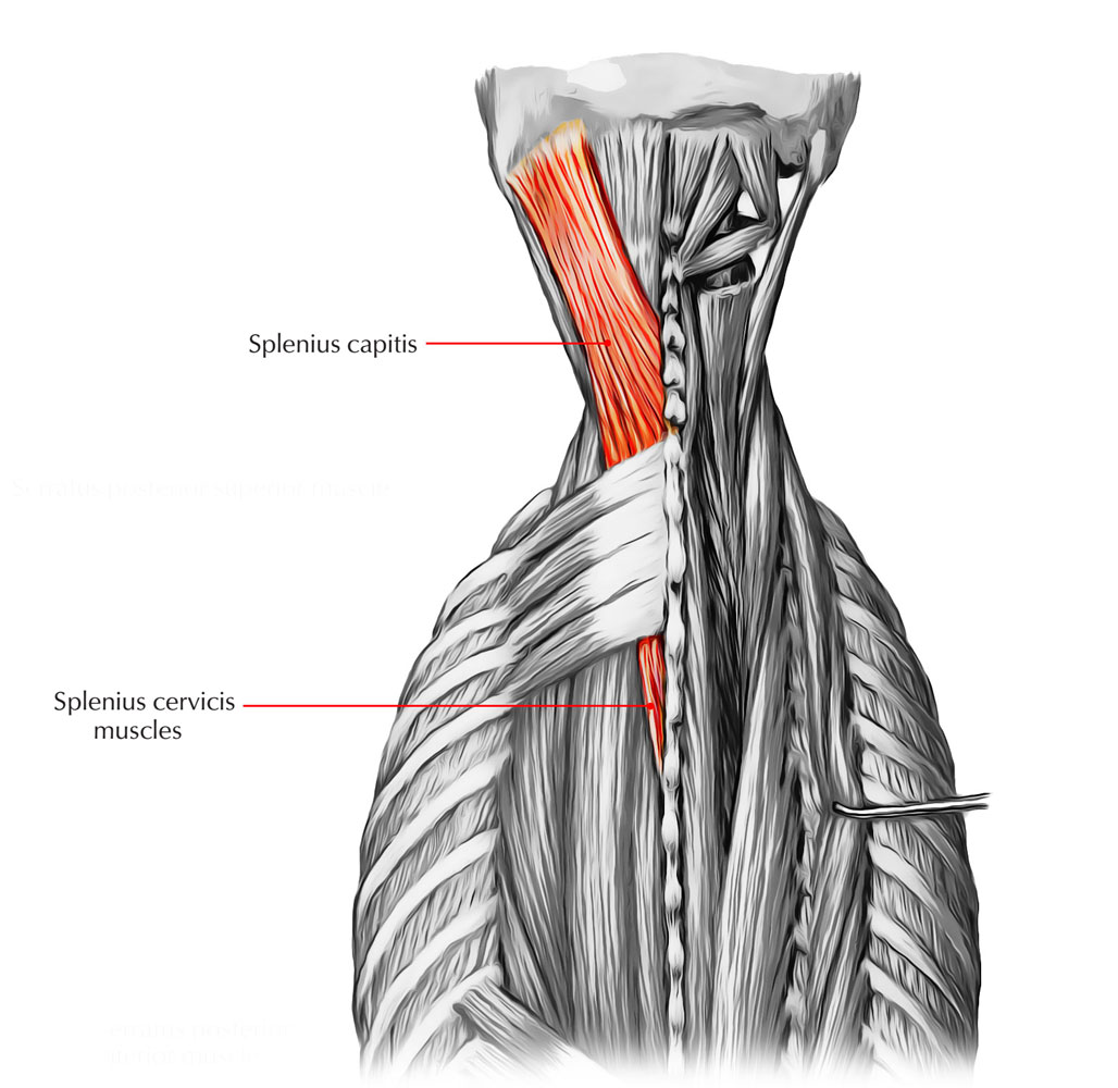
The extrinsic muscles that are associated with upper extremity and thick splenius muscles form the superficial layer of muscles and are located on the lateral and posterior portions of the neck. The spinalis muscle attaches to the spinous processes of the superior thoracic vertebra. The back anatomy includes the latissimus dorsi, trapezius, erector spinae, rhomboid, and the teres significant. The muscles of your back are complex and work together to provide support, movement, and stability. Learn anatomy faster and remember everything you learn.
MRI neck anatomy | free MRI axial neck cross sectional ... from i.pinimg.com Muscles of the back can be divided into superficial, intermediate, and deep group.since the all the back muscles originate in embryo (fetus) form by locations other than the back, muscles in the. This image is titled back muscles ct anatomy and is attached to our article about best back muscles training exercises. Tutorials on the anatomy and actions of the back muscles, using interactive animations, diagrams, and illustrations. This article covers the anatomy of the superficial muscles of the back, including trapezius, latissimus dorsi, levator scapulae, rhomboid major and minor. This is a table of skeletal muscles of the human anatomy. The muscles of the back are a group of strong, paired muscles that lie on the posterior aspect of the trunk. Learn all about the muscles of the back in this 3d video anatomy tutorial. Front view of muscles, skeleton, organs, nervous system.
Here the extrinsic back muscles are classified into logical subgroups to facilitate knowledge.
The extrinsic muscles include the trapezius, latissimus dorsi, rhomboid major and minor, levator scapulae and the serratus posterior superior and. The spinalis muscle attaches to the spinous processes of the superior thoracic vertebra. This is my video about the muscles of the back. Back muscles are divided into two specific groups: The extrinsic muscles that are associated with upper extremity and thick splenius muscles form the superficial layer of muscles and are located on the lateral and posterior portions of the neck. Tutorials on the anatomy and actions of the back muscles, using interactive animations, diagrams, and illustrations. Great round muscle (tere major, tma) little round muscle (teres minor, tmi) infraspinatus muscle (is) these are the main muscle that is responsible for turning and rotating the arm. Muscles of the back can be divided into superficial, intermediate, and deep group.since the all the back muscles originate in embryo (fetus) form by locations other than the back, muscles in the. Fortunately, you don't have to guess. Our back is supported by groups of muscles, which support our posture and ensure stability and balance of the body. Axioappendicular muscles, back muscle basics, back muscles, erector spinae, extrinsic muscles, iliocostalis, intrinsic muscles. These muscles are able to move the upper limb as they originate at the vertebral column and insert onto. Muscle movements, types, and names.
Learn about these muscles, their locations there are several individual muscles within the back anatomy, and it's important to take a quick look at all of them to see how you can target them back muscles anatomy. Back muscles rear view & #8211;

