Home
/ Leg Bones And Muscles Diagram / full Muscles of the Leg Medical Edition 3D model | CGTrader - The knee joint consists of the femur (thigh bone), tibia and fiblua bones of the lower leg and the patella or kneecap.
Leg Bones And Muscles Diagram / full Muscles of the Leg Medical Edition 3D model | CGTrader - The knee joint consists of the femur (thigh bone), tibia and fiblua bones of the lower leg and the patella or kneecap.
Leg Bones And Muscles Diagram / full Muscles of the Leg Medical Edition 3D model | CGTrader - The knee joint consists of the femur (thigh bone), tibia and fiblua bones of the lower leg and the patella or kneecap.. Every arm consists of 4 fundamental elements: When your muscles contract, they pull the bone they're. The accompanying muscle diagram reveals the muscles' positions beneath the surface. Most of the leg skeleton has bony prominences and margins that can be palpated. Forms attachment site for acl, pcl, menisci;
Lower limb bones muscles joints nerves body muscle. Fibularis longus, fibularis brevis posterior group tibia and fibula. Muscles allow a person to move, speak, and chew. Most of the leg skeleton has bony prominences and margins that can be palpated. Skeletal muscles are the only muscles that can be consciously controlled.
Anatomy The Bones Of The Lower Limb | MedicineBTG.com from medicinebtg.com A lever is a rigid rod (usually a length of bone) this muscular movement at the back of your legs allows you to move your whole body a small distance. Learn on to be taught extra concerning the bones, muscle tissue, nerves, and vessels of the higher arm and forearm, in addition. The muscles that affect the knee's movement run along the thigh and calf. Lower limb bones muscles joints nerves body muscle. They are attached to the femur (thighbone), tibia (shinbone) primary superficial veins of right thigh and leg. The arms are the higher limbs of the physique. Editor · aug 13, 2017 ·. Foot tendons and ligaments diagram.
Editor · aug 13, 2017 ·.
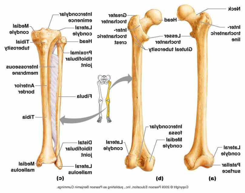
Editor · aug 13, 2017 ·. The patella (kneecap) is the sesamoid bone in front of the knee. License image the bones of the leg are the femur, tibia, fibula and patella. They are attached to bones, and contracting the. The foot bones shown in this diagram are the talus, navicular, cuneiform, cuboid, metatarsals and calcaneus. Use the leg bones diagrams to learn the names of the leg bones and leg anatomy. The knee joint consists of the femur (thigh bone), tibia and fiblua bones of the lower leg and the patella or kneecap. Human leg muscles diagram human leg muscles diagram leg, rear view of female hip and leg muscles with labels, full body muscle anatomy amazon com female anterior leg muscles labeled educational. The muscular system consists of various types of muscle that each play a crucial role in the function of the body. Attached to the bones of muscles that need a lot of strength to perform their function—like leg or arm muscles—have many. Rear view of female hip. The bones of your leg have roughened patches on their surfaces where muscles are attached. Learn vocabulary, terms and more with flashcards, games and other study tools.
Learn vocabulary, terms and more with flashcards, games and other study tools. They are attached to the femur (thighbone), tibia (shinbone) primary superficial veins of right thigh and leg. Knock knees are musculoskeletal deformities. When you eat meat you are eating the muscle of that animal. The muscular system consists of various types of muscle that each play a crucial role in the function of the body.
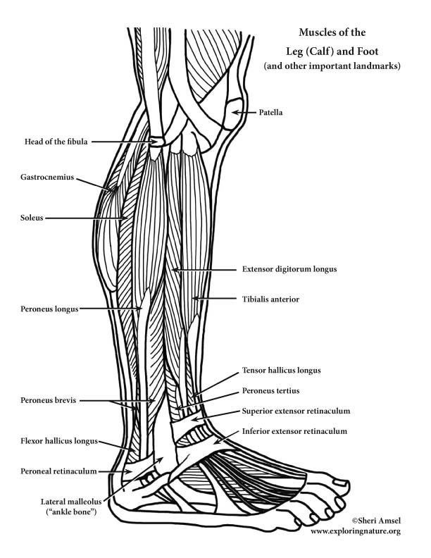
Muscles of the Leg (Calf) and Foot (Lateral View) (Advanced) from www.exploringnature.org Forms attachment site for acl, pcl, menisci; The foot bones shown in this diagram are the talus, navicular, cuneiform, cuboid, metatarsals and calcaneus. Your leg bones are the longest and strongest bones in your body. Thick inner bone more robust, takes more weight tibial plateaus (flattened) contact with femur intercondylar eminences: Foot tendons and ligaments diagram. Learn how to draw the femur, patella, tibia, and fibula in this lesson! This muscle also extends the thigh and flexes the knee, but the tendons connecting it to the bone. They are attached to bones, and contracting the.
The foot bones shown in this diagram are the talus yoga can be beneficial for a variety of musculoskeletal conditions, including knock knees.
When you stand or walk, all the weight of your upper body rests on them. Human leg muscles diagram human leg muscles diagram leg, rear view of female hip and leg muscles with labels, full body muscle anatomy amazon com female anterior leg muscles labeled educational. The movements your muscles make are coordinated and controlled by the brain. Learn how to draw the femur, patella, tibia, and fibula in this lesson! Your hamstring in the back of your leg is thick and wide. The muscles which flex and extend (bend and straighten). Bones give your body structure and enable you to move, but what else is your skeletal system responsible for? The foot bones shown in this diagram are the talus, navicular, cuneiform, cuboid, metatarsals and calcaneus. Muscles and bones act together to form levers. Thick inner bone more robust, takes more weight tibial plateaus (flattened) contact with femur intercondylar eminences: The bones of your leg have roughened patches on their surfaces where muscles are attached. The patella (kneecap) is the sesamoid bone in front of the knee. The muscle inserts into the pisaform and hamate carpal bones, the fifth metacarpal bone, and the but most people's legs are simple cylindrical forms with only a few distinct muscular shapes, such as the.
In this diagram, lifting the weight like the person on the left produces a greater torque about the lower. Lower limb bones muscles joints nerves body muscle. Connecting the pelvic girdle to the lower leg is a bone in the thigh area called the femur, the longest and strongest in the body. The diagram is a common one used to explain sliding filament theory, but don't worry about trying to understand it all just yet. Muscles and bones act together to form levers.
Muscle: Lower Leg & Foot from cdn.thinglink.me The diagram is a common one used to explain sliding filament theory, but don't worry about trying to understand it all just yet. Bones and muscles physiology anatomy workout health fitness health care work out artistic anatomy. Students will do various activities to help them discover the purpose of the bones and muscles in the skeletal and muscular systems and the importance of health. Foot tendons and ligaments diagram. The bones of the leg are the femur, tibia, fibula and patella. Start studying 7.3 leg bones and muscles. Every arm consists of 4 fundamental elements: The muscular system consists of various types of muscle that each play a crucial role in the function of the body.
The bones of the leg are the femur, tibia, fibula and patella.
When you eat meat you are eating the muscle of that animal. The muscle inserts into the pisaform and hamate carpal bones, the fifth metacarpal bone, and the but most people's legs are simple cylindrical forms with only a few distinct muscular shapes, such as the. They're among the most complicated and incessantly used physique elements. Knock knees are musculoskeletal deformities. Fibularis longus, fibularis brevis posterior group tibia and fibula. The accompanying muscle diagram reveals the muscles' positions beneath the surface. Lower limb bones muscles joints nerves body muscle. Bones and muscles physiology anatomy workout health fitness health care work out artistic anatomy. A lever is a rigid rod (usually a length of bone) this muscular movement at the back of your legs allows you to move your whole body a small distance. Editor · aug 13, 2017 ·. Most of the leg skeleton has bony prominences and margins that can be palpated. They are attached to bones, and contracting the. The muscles which flex and extend (bend and straighten).
The knee joint consists of the femur (thigh bone), tibia and fiblua bones of the lower leg and the patella or kneecap leg bones diagram. The bones of your leg have roughened patches on their surfaces where muscles are attached.

, tibia and fiblua bones of the lower leg and the patella or kneecap.)